What Is a Liposuction Scar?
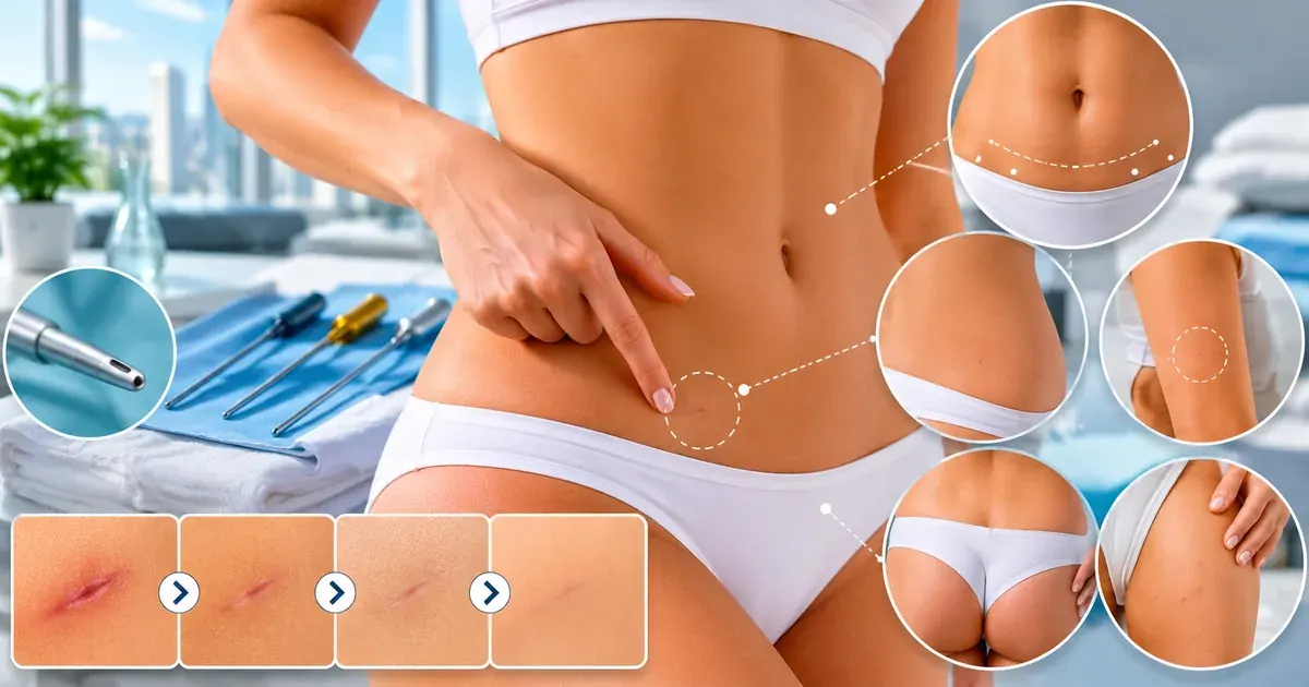
A liposuction scar is not the kind of scar most patients imagine when they hear the word. Unlike open abdominal surgery, a facelift, or a joint replacement — all of which require centimetres-long incisions — liposuction uses only small puncture-style entry points to pass the cannula through. The resulting marks, once healed, look more like a very small freckle or dot than a traditional surgical scar. Most patients are surprised to discover just how inconspicuous their healed incision sites are at 12 months post-surgery.1
That said, a scar is still a scar. Any disruption to the full thickness of the skin — which a liposuction incision is — creates a wound that heals by forming fibrous tissue. The biology is the same whether the incision is 3 mm or 30 cm. The key difference is simply the scale: at 3–5 mm, there is far less surface area for visible scarring, far less tension on the wound edges, and far less opportunity for wider scar formation. Understanding what these marks are — and what they are not — is the first step in managing expectations and taking the steps that genuinely improve outcomes.
It is also important to understand that liposuction scars and post-liposuction fibrosis are two separate phenomena. The scar is the surface mark at the skin puncture site. Fibrosis is a deeper tissue response — firm, lump-like scar tissue in the treated fat layer — that is entirely separate from the visible incision mark. Many patients confuse the two. This guide addresses surface scars in detail; for fibrosis, see the dedicated fibrosis after liposuction guide.
How Incisions Are Made
In tumescent liposuction — the dominant technique used today — the surgeon first infiltrates the target tissue with a large volume of dilute local anaesthetic and adrenaline solution through the same small puncture sites that will be used for cannula access.2 The incision itself is made with a small pointed scalpel (a No.11 or No.15 blade) or a hollow punch, creating a precise circular or slit-like entry point. The width matches the outer diameter of the cannula being used for that area. No tissue is excised; the surrounding skin simply opens to admit the instrument.
Because the incision is created with the anatomy and final scar position in mind from the outset, an experienced surgeon will orient any small slit incision along the natural tension lines of the skin — technically known as Langer's lines or relaxed skin tension lines. An incision cut parallel to these lines heals with less tension and therefore with a finer, less visible scar than one cut perpendicular to them. This is a small technical detail that experienced surgeons apply automatically, but it is worth asking about if you are planning surgery in a region where scarring is a particular concern.
Incision Size by Technique
The diameter of the cannula determines the minimum incision size required. Cannula diameters in common use range from 2 mm to 6 mm, with most standard liposuction performed using 3–4 mm cannulas. Larger-volume areas (abdomen, flanks, outer thighs) often use larger cannulas to work efficiently; finer areas (chin, inner arms, medial knee) use smaller ones. The table below gives a practical overview of typical sizes by technique:
| Technique | Typical cannula diameter | Resulting incision size | Notes |
|---|---|---|---|
| Traditional tumescent liposuction | 3–5 mm | 3–5 mm | Most common technique; suitable for most body areas |
| VASER ultrasound-assisted liposuction | 2.9–4.6 mm probe + cannula | 3–5 mm | Probe inserted first; incision size similar to tumescent |
| Laser liposuction (SmartLipo, SlimLipo) | 1–2 mm fibre + 3–4 mm cannula | 2–5 mm | Laser fibre access may be smaller than aspiration cannula |
| Power-assisted liposuction (PAL) | 3–4 mm | 3–5 mm | Mechanically vibrating cannula; incision size unchanged from tumescent |
| Fine-detail / syringe liposuction | 1.5–2.5 mm | 2–3 mm | Used for face, neck, ankles; smallest incisions |
In practice, the visible difference in healed scar size between a 3 mm and a 5 mm incision is minimal. What matters far more for scar appearance are placement (in a natural crease vs on flat exposed skin), orientation (along vs across skin tension lines), closure technique, and the patient's own healing biology — all covered in detail below.
Where Incisions Are Placed
Surgeons choose incision locations based on two competing priorities: getting the best possible access to the fat being removed, and placing the mark where it will be least visible after healing. In most body areas, experienced surgeons can satisfy both requirements simultaneously by using natural skin creases, body folds, and anatomical landmarks as entry points. The result is that most liposuction scars are hidden in locations that patients — even when looking for them — often cannot find months after surgery.
Placement is also guided by the need for multiple access angles. To produce an even, smooth fat removal without ridges or lumps, the surgeon typically passes the cannula across the same area from at least two different directions. This means two or more incision sites per treated area, though they are all the same small size. On the abdomen, for example, a surgeon might use the navel plus one or two access points in the lower abdominal crease — all hidden by clothing or the natural anatomy.
The table below shows the typical incision locations by body area. Exact placement varies by surgeon preference, patient anatomy, and the specific volume being treated:
| Area treated | Typical incision locations | Why chosen |
|---|---|---|
| Abdomen (upper and lower) | Inside or below the navel rim; lower abdominal hairline crease | Navel scar hidden inside fold; hairline crease covered by underwear |
| Flanks / love handles | Natural back or side fold above or below the waistband line | Hidden by clothing; natural crease reduces scar tension |
| Chin and submental area | Midline just under the chin in the natural crease; behind each ear (if jowls also treated) | Submental crease conceals marks; ear placement hidden by hairline |
| Neck | Submental crease; bilateral below-ear hairline | Natural horizontal crease; hair coverage |
| Upper arms | Axillary (armpit) fold; inner elbow crease | Armpit skin folds hide marks; elbow crease is a natural concealment zone |
| Inner thighs | Inguinal (inner groin) fold; inner knee crease | Groin fold and knee crease rarely visible; covered by most clothing |
| Outer thighs / saddlebags | Below the natural gluteal fold; lateral hip | Gluteal fold conceals well; lateral hip covered by underwear |
| Buttocks / banana fold | Inside the gluteal crease | Deep fold; mark not visible standing or sitting |
| Male chest / gynecomastia | Areola margin (periareolar); axillary fold | Colour change at areola edge camouflages small scar; armpit fold for access |
| Knees / calves / ankles | Posterior knee crease; behind the medial malleolus | Natural flexion crease; bony prominence shadows scar |
Patients should note that the surgeon's first priority when planning incision locations is always safe and effective fat removal. If an area cannot be reached adequately from a hidden location alone, the surgeon will use additional access points that may be more visible. This trade-off is worth discussing at consultation, particularly if you are concerned about specific areas. In most cases, however, the standard hidden-placement approach gives excellent results.
How Lipo Scars Heal: The 4 Phases
Liposuction scars heal through the same four-phase biological process as any wound, but over a longer total timeline than most patients expect. The 3–5 mm incision is small, but the tissue disruption in the underlying fat layer means the local inflammatory environment persists longer than it would for a simple skin cut. Understanding the phases helps patients avoid the common mistake of judging final scar quality too early — particularly at weeks 4–8, when scars are often at their most visible.4
| Phase | Timeframe | What the scar looks like | What is happening biologically |
|---|---|---|---|
| 1. Inflammatory | Days 1–14 | Pink to red; slightly raised; may feel tender or itchy | Blood vessels dilate; immune cells clear debris; haemostasis completed |
| 2. Proliferative | Weeks 2–8 | Can redden further and become slightly raised — often looks worse than week 1; may feel firm | Fibroblasts deposit collagen; new capillaries grow (neovascularisation causes redness); wound closes fully |
| 3. Remodelling | Months 2–18 | Gradually flattens; colour fades from pink/red toward skin tone; softens | Type III collagen replaced by organised Type I collagen; vascularity decreases (redness fades); scar matures |
| 4. Mature scar | 12–18 months+ | Flat, skin-coloured, typically very small and near-invisible | Collagen fully remodelled; no active vascularity; static scar tissue |
Inflammatory Phase: Weeks 1–2
In the first two weeks after surgery, the incision sites are still actively healing. The body sends blood flow and immune cells to the area in a controlled flood — this is the inflammatory phase of wound healing and it is completely normal. The marks will appear pink or light red, may feel slightly warm or itchy, and a small amount of crusting is normal as the wound surface seals. Do not pick at crusts; they are part of the natural closure process and removing them disrupts the healing epidermal layer underneath.
During this phase the incisions may still be covered by your surgeon's dressings, steri-strips, or surgical tape. Follow your surgeon's instructions exactly — the closure support provided by tape reduces the tension on healing wound edges, which directly influences final scar width. At this stage the scar does not yet indicate what it will eventually look like.
Proliferative Phase: Weeks 2–8
The proliferative phase is when fibroblasts — the cells responsible for collagen production — migrate into the healing wound and begin building the structural matrix that will eventually become the scar. This activity requires a rich blood supply, which is why new capillaries grow into the wound (a process called neovascularisation). It is precisely this increased vascularity that makes the scar look redder — and sometimes raised — at weeks 4–8 than it did at week 2. This commonly alarms patients who believe the scar is worsening. It is not: this is the expected appearance of an actively healing wound in full proliferative activity.4
A scar's worst cosmetic appearance during the proliferative phase is not predictive of its final quality. The remodelling phase that follows will transform this temporary dense, vascular tissue into a much quieter, flatter structure. Patients who start scar-care interventions (silicone gel, SPF protection) during this phase — rather than waiting until the scar "looks bad" — achieve better outcomes because they are working with the biology rather than against it.
Remodelling Phase: Months 2–18
The remodelling phase is the longest part of scar healing and the one during which the most dramatic cosmetic improvement occurs. The initial disorganised collagen matrix laid down in the proliferative phase is gradually broken down by enzymes and replaced with more organised, parallel-aligned collagen fibres. As the new vasculature recedes, the redness fades. As the collagen organises, the scar flattens. As the inflammatory stimulus resolves, the firmness softens.
This phase is also when post-inflammatory hyperpigmentation (PIH) is most likely to develop in patients who expose their healing scars to UV radiation. Melanocytes — the pigment-producing cells in the skin — are activated by ultraviolet light in healing tissue, and this activation is far more pronounced in already-inflamed skin. The result is a brown or dark mark at the scar site that can persist for months to years. This is entirely preventable with consistent SPF application and is the single most important scar-care action patients can take. See the scar care section below for detail.
Mature Scar: 12–18 Months
By 12–18 months, the scar has reached its final settled state. Collagen remodelling is essentially complete, the scar is no longer vascularised, and it will not change further without intervention. For the vast majority of patients who had standard liposuction with small cannulas and well-placed incisions, the mature scar is a flat, pale, 3–5 mm mark that is nearly invisible unless specifically searched for. Patients with favourable healing biology, consistent sun protection, and no wound complications often find their scars indistinguishable from normal skin variation at this point.
If a scar at 18 months remains raised, wide, or significantly darker than the surrounding skin, it is worth a consultation with a dermatologist or your original surgeon to assess whether any intervention is appropriate. Options at this stage include silicone-based treatments, pulsed dye laser, fractional laser resurfacing, or intralesional corticosteroid injection depending on the nature of the scar.
Factors That Affect Scarring
Not all patients heal identically. Several biologic, behavioural, and technical factors interact to determine how visible a liposuction scar ultimately becomes. Understanding these factors is not just useful for post-operative management — it helps patients have a more realistic conversation with their surgeon about expected outcomes before surgery, and to identify where they can make a genuine difference to their result through their own behaviour.
The single most impactful factors are largely intrinsic: skin tone, genetics, and age. But several modifiable factors — particularly smoking and sun exposure — can dramatically worsen outcomes for patients who would otherwise heal very well. The combination of a genetic predisposition to darker or raised scarring and poor wound care behaviour can produce a visible scar at a site where better management would have given an invisible one.
Skin Tone and the Fitzpatrick Scale
The Fitzpatrick skin phototype scale classifies skin from Type I (very pale, always burns, never tans) to Type VI (deeply pigmented, never burns). The scale is relevant to scarring because it correlates with melanocyte activity — the skin cells responsible for pigmentation. Patients with higher Fitzpatrick types (IV–VI) have more active melanocytes, which respond more strongly to any inflammatory stimulus, including a healing wound.3
| Fitzpatrick type | Skin description | Post-inflammatory hyperpigmentation risk | Keloid/hypertrophic scar risk |
|---|---|---|---|
| I–II | Very pale to pale; burns easily | Low | Low (keloid very rare) |
| III | Medium; sometimes burns, sometimes tans | Low–moderate | Low |
| IV | Olive; rarely burns, always tans | Moderate–high | Moderate |
| V | Brown; very rarely burns | High | Moderate–high |
| VI | Deeply pigmented; never burns | High | High |
Patients with Fitzpatrick IV–VI skin are not unsuitable for liposuction — the procedure can be performed safely and effectively across all skin types — but they should have an explicit pre-operative conversation with their surgeon about scar risk and the extra vigilance required with sun protection. In some cases, surgeons may recommend prophylactic topical treatments (hydroquinone or azelaic acid) to stabilise melanocyte activity in the peri-operative period, particularly for facial or upper-arm liposuction where the scar is more exposed.
Smoking and Wound Healing
Nicotine is a potent vasoconstrictor — it narrows the small blood vessels (capillaries) that supply oxygen and nutrients to healing tissue. Smokers who undergo surgery have measurably worse wound-healing outcomes than non-smokers across virtually every surgical specialty. The mechanism is well established: reduced capillary perfusion slows every stage of the healing cascade, increases the risk of wound dehiscence (reopening), and results in a wider, thicker scar with greater risk of hypertrophic change.1
Most surgeons advise cessation of smoking for a minimum of four to six weeks before surgery and continuing abstinence for at least six weeks after. The pre-operative period matters because capillary function does not recover instantly after stopping — it takes several weeks for the vasomotor effects of chronic nicotine exposure to reverse. Patients who smoke through recovery, or who resume smoking in the first weeks after surgery, significantly undermine whatever surgical quality their surgeon achieved. For patients who find complete cessation difficult, a nicotine replacement therapy (patch or gum) discussion with their GP before surgery is worthwhile.
Sun Exposure
UV radiation is the single most common preventable cause of conspicuous liposuction scars. Ultraviolet light activates melanocytes in the skin, and in the actively healing, melanocyte-rich environment of a proliferating scar, this activation produces disproportionate pigmentation. The result — post-inflammatory hyperpigmentation — presents as a brown or dark spot at the scar site that can persist for one to several years. In patients with Fitzpatrick IV–VI skin, it can occasionally be permanent without treatment.
The risk is highest during months 2–8, when the scar is in the proliferative and early remodelling phases. The healing tissue has not yet achieved the UV resistance of mature skin, and melanocyte density is temporarily elevated. This is why the standard recommendation — SPF 30 or higher on all incision sites not covered by clothing, for a minimum of six months — is not merely cautious advice. It has a direct biological basis. Patients who cannot reliably apply SPF to incision sites should keep them covered with clothing or opaque tape during outdoor activity in this period.
Surgical Technique
Scar quality is partly a function of the surgeon's choices. Specific technical factors that influence visible scarring include: cannula diameter (smaller produces smaller incision), incision orientation relative to skin tension lines, closure technique (absorbable suture vs steri-strip vs tape), whether incisions are left open for fluid drainage and for how long, and the care taken in achieving a smooth fat removal without excessive trauma to the skin overlying treated areas. Thermal injury to skin edges — possible with laser-assisted liposuction devices if not used carefully — can produce more visible scarring than standard tumescent technique.
When researching surgeons, it is reasonable to ask specifically about their incision placement approach, whether they close incisions or leave them open (and for how long), and what their standard post-operative scar-care protocol is. A surgeon who has a clear, consistent protocol for these details is giving appropriate weight to outcome quality beyond simply the fat removed.
Evidence-Based Scar Care
The scar-care product market is large and not always scientifically rigorous. Patients are frequently confronted with expensive creams, oils, and serums claiming to "eliminate" or "erase" scars — claims that do not reflect how scar biology works. A mature scar is permanently remodelled tissue; no topical product reverses that. What evidence-based scar care can do is modulate the healing process while it is still active, to produce a better final result than no care at all.
The evidence base for specific interventions varies considerably. The strongest evidence is for silicone-based products and for sun protection. The evidence for vitamin E oil, onion extract (Mederma/Contractubex), and most "scar creams" is weak to absent by rigorous clinical standards.3 Patients investing time and money in scar care should prioritise the interventions that have actual data behind them.
Sun Protection
As discussed in the factors section, UV protection of healing incision sites is the most important and most evidence-supported scar-care intervention for the majority of patients. The mechanism is well understood: UV light drives post-inflammatory hyperpigmentation in actively healing tissue. Prevention is straightforward: apply a broad-spectrum SPF 30+ sunscreen to all exposed healing incision sites at least 30 minutes before sun exposure, reapplying every two hours during outdoor activity. Cover with clothing or opaque bandage when sunscreen application is impractical.
This recommendation applies for a minimum of six months from the date of surgery — and longer for patients with darker skin tones who are at higher risk of PIH. Mineral-based sunscreens (zinc oxide, titanium dioxide) are generally preferred over chemical UV filters for healing skin as they are less likely to cause irritation. Do not apply any sunscreen to incompletely closed wounds; wait until the skin surface is fully intact.
Silicone Gel and Sheeting
Silicone-based scar products — available as soft adhesive sheets worn over the scar, or as topical gels applied and left to dry — have the strongest evidence base of any topical scar intervention. Multiple controlled studies and systematic reviews have found that silicone sheeting and gel reduce scar height, improve colour, and decrease symptoms (itch, firmness) compared to controls in patients with hypertrophic and keloid scars.3 The mechanism is thought to involve hydration of the stratum corneum (outer skin layer) and reduction in fibroblast activity through biophysical feedback — silicone maintains a moist, oxygen-rich microenvironment that signals the wound that closure is complete and further collagen deposition is not needed.
Silicone products should only be applied to fully closed wounds — typically from week 2–3 onwards when the surface has sealed. They should be worn or applied consistently: sheets are typically worn 12–23 hours per day; gels are applied twice daily and allowed to dry. Consistency over weeks to months matters more than any single application. Most protocols recommend 2–3 months of use for post-surgical scars, though some surgeons advise continuing for up to 6 months in patients with higher scar risk. Ask your surgeon when to start and which format they recommend; both sheets and gels are clinically comparable in the available evidence.
What Does Not Work
Several popular scar remedies lack meaningful clinical evidence and are worth deprioritising to save both money and time:
- Vitamin E oil: Frequently recommended but poorly supported. A notable randomised controlled trial found no benefit of topical vitamin E over control in post-surgical scar patients, and a significant minority of participants developed contact dermatitis. It is not recommended as a first-line scar treatment.
- Onion extract products (Mederma, Contractubex): Widely marketed and used. Systematic review evidence does not support meaningful benefit over placebo for scar improvement. They are not harmful, but there is no strong reason to prefer them over silicone.
- Cocoa butter and shea butter: Moisturising and well tolerated, these products may prevent excessive dryness around healing wounds, but have no specific scar-modulating evidence. They are fine as a moisturiser; not a substitute for silicone.
- Massage alone (without silicone): Scar massage — gentle circular pressure over a healed scar — is often recommended and may help with scar softening and desensitisation over time. The standalone evidence is limited but it is low-risk and commonly incorporated into post-operative care protocols. It should complement silicone use, not replace it.
- Expensive "stem cell" or "peptide" scar serums: No well-designed clinical evidence supports these products for scar improvement. They are cosmetic, not medical, products and should be evaluated accordingly.
Lumps Near Incisions: Normal vs Concern
One of the most common post-liposuction concerns that patients raise is a firm lump or area of hardness near or under an incision site. Before alarming yourself, it is important to understand that the large majority of such lumps — particularly in the first 2–8 weeks — represent entirely normal healing processes that do not require any intervention. However, some lumps are a signal that something needs assessment. The key is learning to distinguish between them.
The firm hardness felt at the skin surface near an incision site is usually not the surface scar itself. It reflects the underlying tissue response: fibroblast activity, collagen deposition, and local tissue reorganisation in the subcutaneous fat layer where the cannula passed. This is post-liposuction fibrosis — a normal phenomenon covered in detail in the dedicated guide — and it is neither dangerous nor treatment-requiring in the majority of cases. True scar pathology (keloid, hypertrophic scar) affects the skin surface at the incision itself, not the deeper tissue, and presents as a raised, thickened mark at the puncture site rather than a deep lump.
| Finding | Most likely cause | Typical onset | Action needed |
|---|---|---|---|
| Firm, non-tender lump under or near incision | Post-liposuction fibrosis (normal healing) | Days 5–21; peaks weeks 2–6 | None — monitor; resolves over 2–4 months |
| Soft, fluctuant swelling (like a water balloon) | Seroma (fluid collection) | Days 3–21 post-surgery | Contact surgeon — aspiration likely needed |
| Tender, warm, red area near incision | Infection or early haematoma | Days 2–14 (infection), days 1–7 (haematoma) | Contact surgeon promptly — assessment required |
| Raised, thickened scar at incision surface | Hypertrophic scar or early keloid | Weeks 4–12 at incision site | Discuss with surgeon — silicone, steroid injection, or laser |
| Hard, dark nodule within fat layer weeks after surgery | Fat necrosis (rare) | Weeks 2–8 | Contact surgeon for assessment — imaging may be needed |
| Firm area that softens progressively over weeks | Resolving fibrosis (normal) | Weeks 2–12 | None — normal remodelling progress |
The most helpful clinical distinction is texture: fibrosis feels firm and solid, like dense tissue. Seroma feels fluctuant — like a water-filled balloon that shifts when you press it. Infection produces warmth, redness, and tenderness in addition to swelling. Fat necrosis — rare, but possible after aggressive or large-volume liposuction — produces a firm, sometimes irregular nodule within the fat layer that does not behave like resolving fibrosis and may need ultrasound to characterise.
If you are unsure which category a lump falls into, the safest course is to contact your surgeon's office. A lump description over the phone is often enough for them to advise whether you need an in-person assessment or can continue monitoring at home.
Full guide to fibrosis after liposuction →When to Contact Your Surgeon
Most post-liposuction scar and lump concerns are benign and self-resolving. However, specific signs require prompt contact with your surgical team. The following list is not exhaustive, but covers the most common scenarios that warrant medical assessment. When in doubt, contact your surgeon — a brief phone call or message is always appropriate.
Contact your surgeon same day or urgently if you have:
- Fever above 38°C (100.4°F) at any point after leaving hospital
- Increasing redness, warmth, or swelling at an incision site beyond the first 48–72 hours
- Pus or cloudy discharge from an incision site
- An incision that reopens or appears to be separating
- A rapidly growing soft swelling at a treated area (possible expanding seroma or haematoma)
- Severe or increasing pain — post-operative discomfort should be improving from week 2 onwards, not worsening
Contact your surgeon at your next available appointment if you have:
- A lump at an incision site that has not reduced at all by 8–10 weeks
- A raised, thickening scar at an incision site developing beyond weeks 6–8
- Significant darkening or discolouration of the scar site despite sun protection
- Contour irregularities (waviness, dips, ridges) in treated areas at 3–4 months that concern you
- Any symptom that is changing unexpectedly in character — becoming more tender, larger, or different in texture
Persistent scar or contour concerns at 12 months or beyond — after the remodelling phase is fully complete — are a separate category from healing complications. At that point, the question shifts from "is this healing normally?" to "is the final result acceptable, and are there interventions that could improve it?" This is a conversation for a formal surgical review, not an urgent query. Many clinics offer post-operative review consultations at 12 months specifically for this assessment.
Frequently Asked Questions
-
Liposuction does leave scars, but they are extremely small — typically 3–5 mm per incision site. Unlike open surgery, there are no long linear incisions. Surgeons deliberately place these tiny puncture sites in natural skin creases, folds, or areas hidden by clothing. For most patients, fully healed lipo scars are nearly undetectable to the naked eye at 12–18 months after surgery.
-
Technically yes — any healed wound leaves some degree of permanent mark. In practice, liposuction incision scars are so small (3–5 mm) and placed so strategically that most patients consider them negligible after 12–18 months. The scar typically fades from pink or red to skin-coloured during the remodelling phase. Patients with darker skin tones (Fitzpatrick IV–VI) or a genetic history of keloid scarring may retain more visible scars long-term.
-
The most impactful single step is protecting incision sites from sun exposure for at least 6 months — ultraviolet radiation causes post-inflammatory hyperpigmentation that permanently darkens healing scars. Other evidence-supported measures include applying silicone gel or sheeting to fully closed wounds, avoiding smoking throughout the healing period, keeping wounds moisturised once closed, and not picking or stretching healing tissue. Follow your surgeon's specific wound-care protocol, as instructions vary by technique.
-
A firm, non-tender lump near or under an incision site at weeks 2–8 after liposuction is most likely fibrosis — normal scar tissue formation in the underlying treated tissue. This is distinct from the surface scar and resolves naturally over 2–4 months in the majority of patients without any intervention. A lump that is tender, warm, growing, or accompanied by fever or skin redness may indicate a seroma or infection and should be assessed by your surgeon promptly.
-
Keloid or hypertrophic scarring is possible after liposuction, though uncommon given the very small incision sizes. Patients with a personal or family history of raised, widening scars are at higher risk. If you have this history, discuss it with your surgeon before the procedure — incision placement, closure technique, and post-operative scar management may be modified. Early treatment of any raised scar with silicone sheeting or corticosteroid injections is most effective when started promptly.
-
The full scar maturation process takes 12–18 months. Scars are typically most visible — and often at their worst in terms of redness and slight elevation — at weeks 4–8 during the proliferative healing phase. This is normal and not a predictor of final outcome. From months 2 onwards, scars gradually flatten and fade through the remodelling phase, reaching their final skin-coloured, near-flat appearance by 12–18 months for most patients.